Brain Scans
Note: Remember to click on any word on this page to experience the next evolutionary step in technology supported reading.
What Brain Scans Reveal
Well, clearly, for anyone who’s interested in reading and dyslexia, which was initially described over a hundred years ago, the goal has been to try to understand where, at a fundamental level, reading takes place; and obviously it takes place in the brain. However, the challenge has been: How do you study a process within the brain of children who are otherwise healthy? Fortunately, we have a very nice hard skull protecting the brain. So how do you look within the brains of healthy children? We’ve all been extraordinarily fortunate that two lines of research have converged. One is the understanding of the basic nature of reading and reading difficulties; and the other has been the extraordinary progress in technology that allows us in really quite a benign way to be able to image the brain at work. We use Functional Magnetic Resonance Imaging (fMRI), which allows us to image children and adults as they try to read without using radiation or injections or anything like that.
So, our first goal was to see if the technology could work, and to be able to identify and localize what are the brain systems used in reading. First we studied adults, good and poor readers. We found that good readers use three major systems on the left side of the brain. Then we found that poor readers had a disruption, a significant underactivation of two of the regions in the back of the brain. But when you see that, the question always is: Is this something that if you see it in an adult, is it the result of years and years of not reading?
So, our next step was to study a very, very large group of children who were good readers and struggling readers, and we found exactly what we had found in the adults, a significant underactivation in the areas in the back of the brain. This study was really a very, very informative one because there was an unusually large number of children participating, 144. That is a very large number of children for an imaging study and that allowed us to do a number of things. It allowed us to begin to map out: Well, what do these areas do? What was particularly of interest to us was one of the areas in the back of the brain, the left occipital temporal region, or the word forming area. Activation in that area was significantly related to a child’s skill as a reader.
So the individual differences in reading skills were related to individual differences in brain activation. That was very important, because this is a very important area in fluency, which is critical to being able to read not only accurately, but also rapidly and with good intonation. So, we went from simply geographically identifying the systems to being able to see the locations of potential differences between good and poor readers, and what the role of each of these systems is.
Sally Shaywitz, Pediatric Neuroscience, Yale University, Author of Overcoming Dyslexia. Source: COTC Interview – http://www.childrenofthecode.org/interviews/shaywitz.htm#WhatBrainScans
Limitations of Current Brain Scans
David Boulton: Do we have any brain imaging tests that you’re aware of that allow us to see a signature distinction in the structure or processing of the brain before the process of learning to read that predicts difficulty in learning to read?
Dr. Sally Shaywitz: We don’t. I know exactly what you’re saying, and that would be just a really commendable goal. Currently, when we do functional brain imaging, children have to lie very still, and the youngest that I think we or others have done is about six-and-a-half. So then at that time that would be when children are about near the end of kindergarten. So you’d expect them to have the rudiments of early reading in place.
It may be possible, with other technologies that are evolving, to be able to identify children earlier.
David Boulton: So, currently there’s a rather significantly sized uncertainty about that threshold.
Dr. Sally Shaywitz: That’s right.
David Boulton: Okay. Relative to your tests, how fast a frequency variation can you see? I mean, for example, with Keith Rayner’s work we’re seeing that the difference between graphemic and phonological follow-up is happening in a five to twenty-five millisecond time window when somebody is reading well.
Dr. Sally Shaywitz: Well, Functional Magnetic Resonance Imagining doesn’t give temporal information.
David Boulton: So, then difficulties associated with the time precarious interactive interplay between these various modules you’re identifying can’t be seen with the equipment and setups we’ve got yet.
Dr. Sally Shaywitz: Certainly not with the Functional MRI. But what we can see, for example, that when we present words to children very quickly if they are using the word formation area that’s critical for fluent reading. That’s possible.
Now there are new techniques that show how the parts of the brain are connected, so we can have a sense — and that’s what I was just telling you about in the last research I mentioned, where we looked at the persistently poor readers who seemed to have all the systems in place, but they weren’t connected appropriately, they weren’t connected properly. I think this is an area where the technology is evolving very, very rapidly, so I think things are really changing as we speak.
David Boulton: So, we’re heading towards being able to peer into this space. It seems when children are reading, if we really track their facial expressions and articulations, there’s a hesitancy, or starting and stopping that happens for struggling readers, and that that starting and stopping has a correspondence to a number of things. One thing for sure is the kind of ambiguity they’re processing at that moment, yes?
Dr. Sally Shaywitz: Well, yes, I think each of us would represent that a little bit differently.
Sally Shaywitz, Pediatric Neuroscience, Yale University, Author of Overcoming Dyslexia. Source: COTC Interview – http://www.childrenofthecode.org/interviews/shaywitz.htm#LimitationsofCurrentBrain
Magnetic Imaging Methods
Because we now have the advantage of technological imaging in non-invasive imaging in humans both through evoked potential electro-physiological methods and also through functional neuro-imaging methods. Both magnetic imaging methods have been able to show that, indeed, not only are the children behaviorally processing more quickly, their language and reading scores on standardized tests are improving. And the differences between their brains and the brains of children who are better readers are becoming less distinct over time. So, those areas that are more involved in certain aspects of the reading task, particularly the phonological aspects, which are distinctly different between a group of children who are struggling with reading and a group of children who are reading really well are diminishing.Over time, if your methodologies of intervention are working, one should be able to determine that using not only behavioral methodologies works, but also physiological and neuro-imaging. That’s a real advance. This is brand new data. For the first time we’re beginning to see studies in which one is able to actually evaluate the effectiveness of reading interventions or other types of interventions. Very similar kinds of approaches are being used with patients with stroke, both for the motor systems, the language systems, and the perceptual system and sensory systems. Particularly, we’ll see more and more studies coming out in which these neuro-imaging procedures are being used as assessments for the efficacy; first of all assessments for a deficit, and then used as an additional way of evaluating efficacy of one method over another. It’s a new world.
Paula Tallal. Board of Governor’s Chair of Neuroscience and Co-Director of the Center for Molecular and Behavioral Neuroscience at Rutgers University. Source: COTC Interview – http://www.childrenofthecode.org/interviews/tallal.htm#ExercisesToSpeedUpDistinctionProces
Newer Technologies Image the Brain in Real Time
The reality is that if you look at these newer technologies that are able to image the brain in real time – not just take a picture of what lit up and subtract it out from everything else, but really follow using full head evoked potential or magnetoencephalography or something that can actually follow the response – in general, a lot of the brain is working to process information as complex as speech. And when you get into reading where you’ve got the language plus the written part to be decoded and reassembled and comprehended, we are really using a lot of parts of our brain.
Paula Tallal. Board of Governor’s Chair of Neuroscience and Co-Director of the Center for Molecular and Behavioral Neuroscience at Rutgers University. Source: COTC Interview –http://www.childrenofthecode.org/interviews/tallal.htm#AmbiguityProcessingTakesTime
Limitations of Current Brain Imaging Technologies
David Boulton: We’ve talked to neuroscientists who are studying the brain at different stages in the reading process. And they’re basically saying variations of what you said in your article regarding the limitations of current brain imagining technology. We aren’t yet able to peer into the levels of details and at the rate of processing that this construction is occurring in.
Dr. Christof Koch: Yes, we have EEG that of course can resolve the timing, but that doesn’t allow us to really pinpoint anything, because it doesn’t really allow us to pinpoint the signals in terms of particular parts of the brain. We’ve got an fMRI, which allows us to pinpoint, this part of the brain or that part of the brain, but it’s very sluggish because it works on a time scale of many seconds.
David Boulton: Right. I make the analogy; it’s like a satellite looking at the aggregate light patterns of Los Angeles and Houston at night. We can see the difference in the intensity of light between the two cities, but we can’t see the federal express messages going back and forth between them.
Dr. Christof Koch: No, no.
David Boulton: Well…
Dr. Christof Koch: That’s what we’re stuck with right now. We have more options in the study of animals, but of course, no animal is able to read
Christof Koch, Professor of Cognitive and Behavioral Biology, California Institute of Technology. Director, Koch Laboratory, member of the Division of Biology as well as the Division of Engineering and Applied Science. Source: COTC Interview: http://www.childrenofthecode.org/interviews/koch.htm#Limitations_of_Current_Brain_Imaging_Technologies
BUY CHILDREN OF THE CODE VIDEOS:
THE FUTURE OF LEARNING TO READ AND READING TO LEARN:
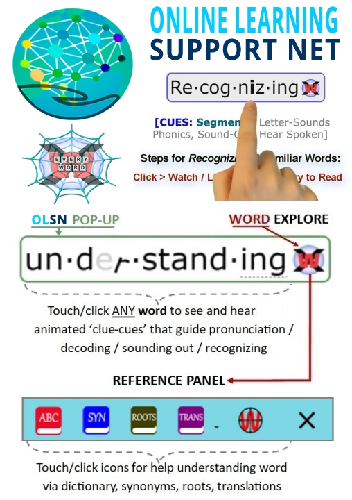
FREE K-ADULT LIBRARY FULL OF OLSN ENABLED STORIES AND GAMES:
INSTALL FREE OLSN CHROME EXTENSION:

-------
PLEASE HELP US
Learning Stewards is a 501(c)(3) non-profit that depends on donations from people like you. Please donate whatever you can.
Turn your Amazon shopping into support for the Children of the Code Project and help us help children, parents, and educators!